


PD-1
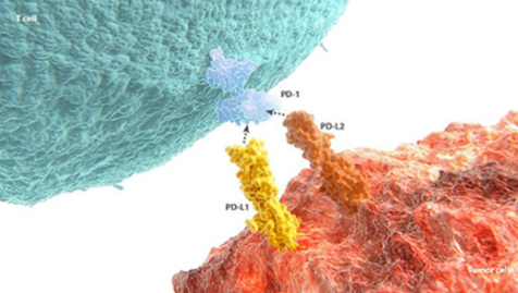
免疫チェックポイント受容体であるPD-1(Programmed death receptor-1)は、腫瘍環境で過剰発現しており、T細胞の疲弊化が促進されるため、抗腫瘍免疫応答が減弱します。PD-1の阻害は細胞傷害性T細胞  の免疫機能を賦活化させます。
発現および生理的な機能
- PD-1は細胞傷害性T細胞上に発現する免疫チェックポイント受容体です。
PD-1のリガンドには、PD-L1(Programmed death ligand 1)およびPD-L2(Programmed death ligand 2)の2つが存在します1-3。
- これらのリガンドは、T細胞の活性を阻害する点において、機能が重複しています1,2。 - PD-1およびPD-L1、またはPD-1およびPD-L2の発現上昇は、T細胞の疲弊化に重要な役割を果たしますが、一方では自己免疫の回避において重要な因子ともなります3-5。
がんでの役割
- 腫瘍では、PD-L1およびPD-L2ががん細胞の表面に発現し、T細胞を疲弊させます1-3,6-8。
- これは、がんの免疫からの回避におけるPD-L1およびPD-L2の役割を示唆しています9-12。
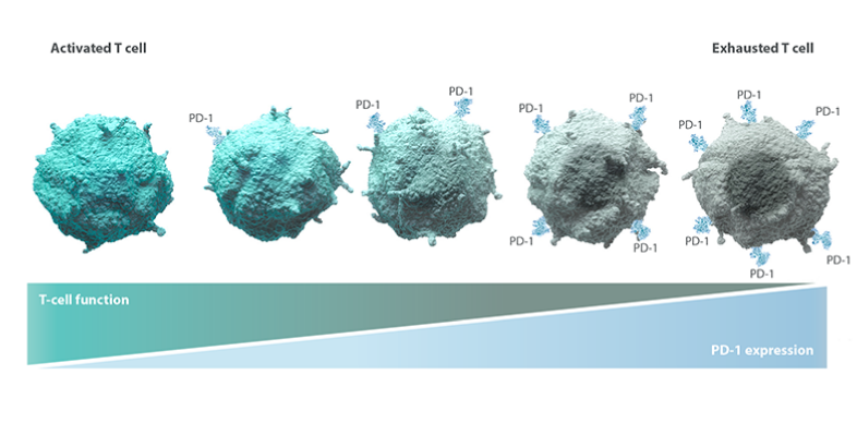
- 実臨床のデータでは、PD-L1およびPD-L2が、腎細胞癌、悪性黒色腫、非小細胞肺癌、食道癌、膵臓癌、肝細胞癌、リンパ腫を含む、複数の固形がんおよび造血器腫瘍で発現していることが示されています8-13。 - 腫瘍微小環境では、がん抗原への反復的な曝露によりPD-1活性が次第に増強し、T細胞の疲弊化が進行します。
つまり、PD-1シグナル伝達が制限無しに増加することで、T細胞の免疫応答機能が失われることになります3,6,7。
- T細胞は、疲弊化の進行により、増殖能力、がん細胞を攻撃する能力、そして最終的に生存能力といった、必須機能を失っていきます14。 - 固形がんおよび造血器腫瘍における腫瘍浸潤T細胞は、しばしば次のような疲弊の証拠を示します。
- PD-1および他の免疫抑制因子の発現上昇3
- 免疫応答を誘導する細胞シグナル伝達分子(サイトカイン)の減少3,15
- がん細胞を攻撃する能力の低下3,15
前臨床のエビデンス
- 前臨床試験では、PD-1阻害により疲弊したT細胞が再活性化し、細胞傷害性免疫機能が回復することが示されています16。
- さらに、前臨床試験では、PD-L1およびPD-L2の両者を阻害し、PD-1シグナル伝達を完全に阻害すると、PD-L1単独を阻害するときよりも、疲弊したT細胞を再活性化する効果は大きいことが示唆されています17。 - PD-1経路の阻害により弊したT細胞の再活性化に対する影響、およびPD-1阻害と他の免疫経路との関連性についての解明を目指した研究が進められています。
REFERENCES–PD-1
- Freeman GJ, Long AJ, Iwai Y, et al. Engagement of the PD-1 immunoinhibitory receptor by a novel B7 family member leads to negative regulation of lymphocyte activation. J Exp Med. 2000;192(7):1027-1034.
- Latchman Y, Wood CR, Chernova T, et al. PD-L2 is a second ligand for PD-1 and inhibits T cell activation. Nat Immunol. 2001;2(3):261-268.
- Ahmadzadeh M, Johnson LA, Heemskerk B, et al. Tumor antigen–specific CD8 T cells infiltrating the tumor express high levels of PD-1 and are functionally impaired. Blood. 2009;114(8):1537-1544.
- Barber DL, Wherry EJ, Masopust D, et al. Restoring function in exhausted CD8 T cells during chronic viral infection. Nature. 2006;439(7077):682-687.
- Nishimura H, Okazaki T, Tanaka Y, et al. Autoimmune dilated cardiomyopathy in PD-1 receptor-deficient mice. Science. 2001;291(5502):319-322.
- Catakovic K, Klieser E, Neureiter D, Geisberger R. T cell exhaustion: from pathophysiological basics to tumor immunotherapy. Cell Commun Signal. 2017;15(1):1. doi:10.1186/s12964-016-0160-z.
- Peng W, Liu C, Xu C, et al. PD-1 blockade enhances T-cell migration to tumors by elevating IFN-γ inducible chemokines. Cancer Res. 2012;72(20):5209-5218.
- Taube JM, Klein A, Brahmer JR, et al. Association of PD-1, PD-1 ligands, and other features of the tumor immune microenvironment with response to anti–PD-1 therapy. Clin Cancer Res. 2014;20(19):5064-5074.
- Nomi T, Sho M, Akahori T, et al. Clinical significance and therapeutic potential of the programmed death-1 ligand/programmed death-1 pathway in human pancreatic cancer. Clin Cancer Res. 2007;13(7):2151-2157.
- Ohigashi Y, Sho M, Yamada Y, et al. Clinical significance of programmed death-1 ligand-1 and programmed death-1 ligand-2 expression in human esophageal cancer. Clin Cancer Res. 2005;11(8):2947-2953.
- Hamanishi J, Mandai M, Iwasaki M, et al. Programmed cell death 1 ligand 1 and tumor-infiltrating CD8+ T lymphocytes are prognostic factors of human ovarian cancer. Proc Natl Acad Sci U S A. 2007;104(9):3360-3365.
- Green MR, Monti S, Rodig SJ, et al. Integrative analysis reveals selective 9p24.1 amplification, increased PD-1 ligand expression, and further induction via JAK2 in nodular sclerosing Hodgkin lymphoma and primary mediastinal large B-cell lymphoma. Blood. 2010;116(17):3268-3377.
- Jung HI, Jeong D, Ji S, et al. Overexpression of PD-L1 and PD-L2 is associated with poor prognosis in patients with hepatocellular carcinoma. Cancer Res Treat. 2017;49(1):246-254.
- Lee J, Ahn E, Kissick HT, Ahmed R. Reinvigorating exhausted T cells by blockade of the PD-1 pathway. For Immunopathol Dis Therap. 2015;6(1-2):7-17.
- Zhang L, Gajewski TF, Kline J. PD-1/PD-L1 interactions inhibit antitumor immune responses in a murine acute myeloid leukemia model. Blood. 2009;114(8):1545-1552.
- Sznol M, Chen L. Antagonist antibodies to PD-1 and B7-H1 (PD-L1) in the treatment of advanced human cancer. Clin Cancer Res. 2013;19(5):1021-1034.
- Hobo W, Maas F, Adisty N, et al. siRNA silencing of PD-L1 and PD-L2 on dendritic cells augments expansion and function of minor histocompatibility antigen–specific CD8+ T cells. Blood. 2010;116(22):4501-4511.


ブリストル・マイヤーズ スクイブ株式会社
米国本社サイト
このサイトは、日本国内の医療関係者の方を対象にブリストル・マイヤーズ スクイブ株式会社の医療用医薬品を適正にご使用いただくために作成したものです。